What Is a Ventricular Septal Defect?
The normal heart has four chambers: two upper chambers called atria, which receive blood into the heart, and two lower chambers called ventricles, which pump blood out of the heart to the lungs or the rest of the body. Ordinarily, there is a wall of muscle between the heart’s chambers that separates them completely. A ventricular septal defect (VSD) is one or more holes in the wall (called the ventricular septum) between the heart’s main pumping chambers, or ventricles.
These holes cause blood to flow from the left ventricle into the right ventricle and into the lungs. The extra blood in the lungs overworks the lungs and the heart.
VSD is the third most common birth defect in Texas, with 5.6 cases for every 1,000 live births, according to the latest research from the Texas Birth Defects Registry of data through 2011.
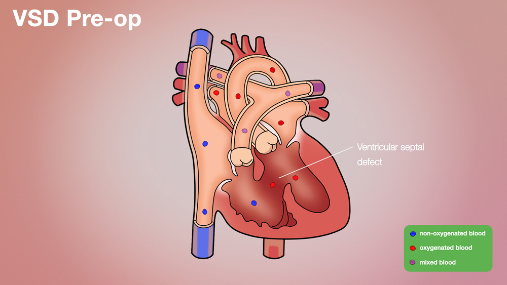
Ventricular Septal Defect (VSD) Pre-op
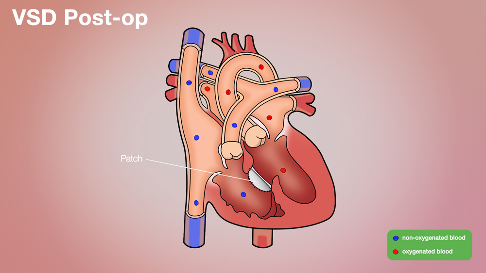
Ventricular Septal Defect (VSD) Post-op
The holes have different names, depending on their location:
- Conoventricular VSD refers to a hole just below the pulmonary aortic valves.
- Perimembranous VSD is a hole in the upper section of the septum.
- Inlet VSD is a hole near where the blood enters the ventricles, via the tricuspid and mitral valves. This defect may be part of another condition called an atrioventricular septal defect.
- Muscular VSD, the most common VSD, is a hole in the lower, muscular part of the wall.
What Are the Causes?
VSD is a congenital birth defect that occurs when the wall between the heart’s lower chambers does not fully develop as the heart forms during the first eight weeks of pregnancy. Genes may be involved: If you have a child with a congenital cardiac defect, the chance of having another child with a heart defect is about 2% to 3%. This may depend on the type of heart defect your child has.
How Is It Diagnosed?
VSD may be diagnosed during pregnancy with a fetal echocardiogram, which is a specialized ultrasound of the fetal heart. The affiliated physicians in the Fetal Cardiology Program at The Fetal Center will confirm a diagnosis and prepare a delivery plan for both mom and baby. A multidisciplinary team of specialists will also develop the baby's immediate care plan following delivery.
If a VSD is not diagnosed in utero, and suspicion of a heart defect occurs after the baby is born, a pediatrician will refer the patient to a pediatric cardiologist to determine the diagnosis.
If the hole is small, it usually closes on its own. The only sign may be a heart murmur – or whooshing sound – that the physician may hear via a stethoscope.
If the hole is large, side effects may be noted in newborns or may not appear until months later. Signs include short, fast or heavy breathing; sweating and/or fatigue while feeding; and poor weight gain. Over time, the overworked heart and lungs may lead to erratic heartbeats, heart failure, or high blood pressure in the lungs (pulmonary hypertension).
The following tests may be utilized to find more details:
- A cardiac echocardiogram (echo), using sound waves (ultrasound) to produce images of the heart and blood vessels on a screen. This painless exam will show structural problems, the size of the hole in the ventricular wall and how much blood is flowing through it. The echo also reveals whether too much or not enough blood is going to the lungs or the body as a whole.
- Chest X-rays, which create images of the heart and lungs, making major flaws visible, such as enlargement of the heart or fluid buildup in the lungs.
- An electrocardiogram (EKG or ECG) is a painless exam that evaluates the heart’s electrical system to reveal irregular rhythms, suggesting a stressed heart.
- A cardiac MRI (magnetic resonance imaging) is a painless exam using radio waves, magnets and a computer to form three-dimensional images of the heart, which can reveal holes in the ventricular wall and other structural abnormalities.
- Cardiac catheterization, which involves a thin, long tube that is inserted into a blood vessel at the groin and guided via an artery or vein into the heart. The doctor can see more details of the hole in the ventricular wall and determine blood pressure and oxygen levels in the four heart chambers and surrounding blood vessels.
How Is It Treated?
The child’s pediatric cardiologist will monitor him or her to see whether the hole, if small, closes on its own. Medication cannot close a hole, but it can help with related issues caused by the defect, by lowering blood pressure, strengthening the heart muscle, or ridding the lungs and body of excess fluid (via diuretics). A baby who is failing to gain weight might be fed a high-calorie formula or fed via a feeding tube.
Depending on the hole’s size and the child’s symptoms, there are three ways that might be used to close a VSD.
If appropriate, doctors may perform a cardiac catheterization to close the hole. Specially trained cardiologists insert a device into one of the large veins in the groin. A closure device is delivered in collapsed form through the vein to the site of the hole in the heart. It then can be expanded and placed into position to close the hole. In order to be closed with this minimally invasive technique, gaps must be surrounded by enough tissue to seat the device.
In very small children, or when catheter closure is difficult, a hybrid, or collaborative procedure between an interventional cardiologist and a heart surgeon, may take place where a surgery is performed, but the defect is closed with a catheter-based device without the use of a heart-lung bypass machine. The ability to close VSDs with a device is mostly depended on their location in the ventricular septum, and whether the device will affect the heart valves or electrical system.
The third option for VSD closure involves open heart surgery. Surgery is usually performed through a vertical (up and down) incision in the middle of the chest. Repair does require open heart surgery and the patient is put on the heart-lung bypass machine. The machine oxygenates the blood and then pumps it back into the body. In this way, the surgeon can work inside the heart easily.
Surgical closure of the hole involves a patch of man-made material or the outside lining of the heart (pericardium) to cover the hole. The patch is secured to the edge of the hole using surgical sutures. Since the VSD is in the middle of the heart, a small incision is made in the right atrium to gain access to the VSD; the incision poses no damage to the heart.
What Are the Long-Term Effects?
Recovery after a VSD closure varies with age. Children usually spend anywhere from 3-8 days in the hospital. Babies and younger children may require longer hospitalization and recovery time, depending on the health of their lungs and their ability to eat and gain weight. If a school-age child has a VSD repair, he or she will typically be out of school for 3-4 weeks after surgery for recovery.
Over time, the natural lining of the heart usually grows to cover any patch, and the repair will become permanent. The child’s pediatric cardiologist will check regularly for the rare potential complication of a leaky valve.
The doctor also may prescribe antibiotics before any dental procedures in the six months following surgery, to prevent inflammation of the heart’s inner lining (endocarditis). Uncommonly, high blood pressure will arise in the lung’s vessels (pulmonary hypertension), and medication will be prescribed. Usually no follow-up surgery or medications are needed.
What Follow-Up Care Is Needed?
If the VSD was small or was closed in childhood, usually checkups every three to five years are sufficient, reports the American Heart Association. If the VSD was large, checkups will be more frequent.
Why Choose the Children’s Heart Institute?
At Children’s Heart Institute at Children’s Memorial Hermann Hospital, patients with congenital or acquired heart disorders receive hands-on specialized care 24/7 from a team of affiliated physicians and specialty-trained nurses who aim to deliver the best possible outcomes.
Children’s Memorial Hermann Hospital was named one of the top children's hospitals nationally in Cardiology & Heart Surgery by U.S. News & World Report. In addition, Children’s Heart Institute is among the top congenital heart surgery programs in North America for patient care and outcomes, according to the Fall 2019 Society of Thoracic Surgeons (STS) Congenital Heart Surgery Database Report of 118 STS participating programs.
In collaboration with various subspecialties, the affiliated team provides comprehensive care for newborns, children and adolescents, with the ability to transition into adult congenital cardiac care. Team members have the experience and skills necessary to offer innovative treatment methods and specialized services, including, but not limited to:
- Biventricular repairs and biventricular conversions
- Congenital heart optimization
- Full repairs for complex congenital heart defects in newborns
- Hybrid catheterization and surgical procedures
- Minimally invasive transcatheter pulmonary valve (TPV) therapy
- Minimally invasive repairs
- Treatment for adult congenital heart disease
- Valve repairs and preservation
With the Level IV Neonatal Intensive Care Unit (NICU) and a dedicated Children’s Heart Institute Intensive Care Unit at Children’s Memorial Hermann Hospital, critical heart patients have access to quality, specialized care. By utilizing state-of-the-art techniques, the team at Children’s Heart Institute strives to offer patients with the most complex problems the greatest opportunity for a normal life.
Contact Us
If you have any questions, use the online tool below to help us connect with you. To refer a patient or schedule an appointment, please contact our clinic using the information below.
- Pediatric Cardiology Clinic
The University of Texas Health Science Center Professional Building
6410 Fannin, Suite 370
Houston, TX 77030
Phone: (713) 486-6755 (Appointment Line)
- Pediatric and Congenital Heart Surgery Clinic
The University of Texas Health Science Center Professional Building
6410 Fannin, Suite 370
Houston, TX 77030
Phone: (713) 500-5746
CMHH-Heart@memorialhermann.org
To contact Children’s Heart Institute at Children’s Memorial Hermann Hospital, please fill out the form below.
The Children’s Heart Institute is a collaboration between the affiliated physicians at McGovern Medical School at UTHealth Houston and Children’s Memorial Hermann Hospital. Typically, patients are seen on an outpatient basis at a UT Physicians clinic with all inpatient procedures performed at Children’s Memorial Hermann Hospital.
Nationally Ranked Pediatric Care
Proud to be named one of the nation’s best in Cardiology and Heart Surgery; Gastroenterology and GI Surgery; Neonatology; and Neurology and Neurosurgery.
Learn More